Home

Presunzione conformarsi ponte jc 1 staining protocol Insistere esaminare schema

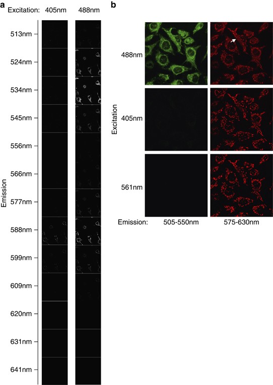
JC-1: alternative excitation wavelengths facilitate mitochondrial membrane  potential cytometry | Cell Death & Disease
JC-1: alternative excitation wavelengths facilitate mitochondrial membrane potential cytometry | Cell Death & Disease
JC-1 Experiment Common Questions and Solutions

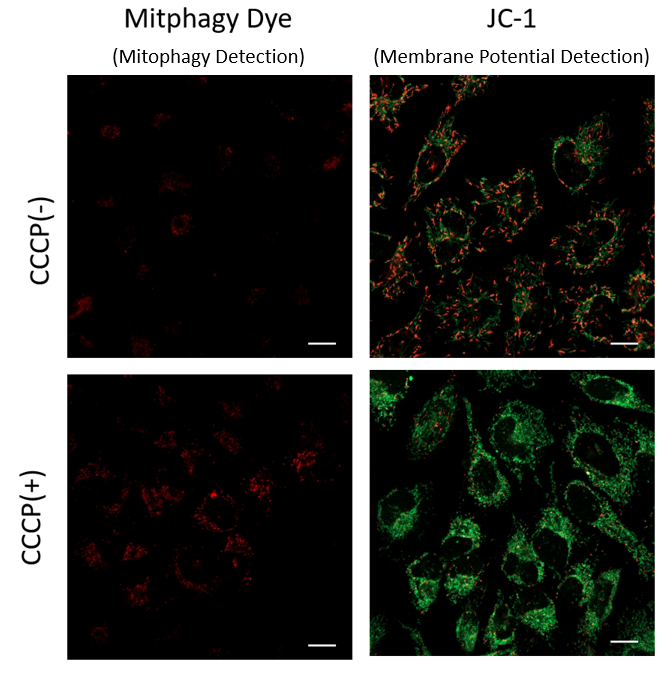
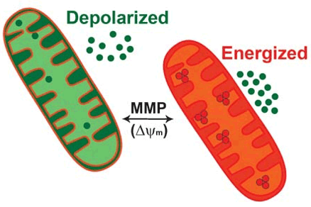
Mitochondrial Membrane Potential Detection JC-1 MitoMP Detection Kit Dojindo

HY-15534-5mg | JC-1 [3520-43-2] Biotrend

Figure 1. JC-1 staining of peripheral blood lymphocytes and monocytes. Note  the different fluorescence intensity of the two cell types, due to the  presence of a higher number of mitochondria in monocytes.
Figure 1. JC-1 staining of peripheral blood lymphocytes and monocytes. Note the different fluorescence intensity of the two cell types, due to the presence of a higher number of mitochondria in monocytes.

HY-15534-5mg | JC-1 [3520-43-2] Biotrend

www.bio-protocol.org/e3128 Analysis of the Mitochondrial Membrane Potential  Using the Cationic JC-1 Dye as a Sensitive Fluoresce
www.bio-protocol.org/e3128 Analysis of the Mitochondrial Membrane Potential Using the Cationic JC-1 Dye as a Sensitive Fluoresce

JC-1 Mitochondrial Membrane Potential Assay | Creative Bioarray

Detection of mitochondrial membrane potential by JC-1 staining after... |  Download Scientific Diagram
Detection of mitochondrial membrane potential by JC-1 staining after... | Download Scientific Diagram

JC-1: alternative excitation wavelengths facilitate mitochondrial membrane  potential cytometry | Cell Death & Disease
JC-1: alternative excitation wavelengths facilitate mitochondrial membrane potential cytometry | Cell Death & Disease
1.2 Vitexin and the Mitochondria – Selected Topics in Health and Disease  (2019 Edition)
1.2 Vitexin and the Mitochondria – Selected Topics in Health and Disease (2019 Edition)

HY-15534-5mg | JC-1 [3520-43-2] Biotrend

JC-1 - Mitochondrial Membrane Potential Assay Kit (ab113850) | Abcam

Mitochondrial Membrane Potential and Apoptosis Assay Kit

Time-lapse imaging of JC-1-positive cells. (A): The JC-1... | Download  Scientific Diagram
Time-lapse imaging of JC-1-positive cells. (A): The JC-1... | Download Scientific Diagram

JC-1 Dye for Mitochondrial Membrane Potential | Thermo Fisher Scientific -  TW
JC-1 Dye for Mitochondrial Membrane Potential | Thermo Fisher Scientific - TW

JC-1
JC-1

JC-1 (CBIC2) | Mitochondrial Membrane Potential Probe | MedChemExpress

JC-1 - Mitochondrial Membrane Potential Assay Kit (ab113850) | Abcam

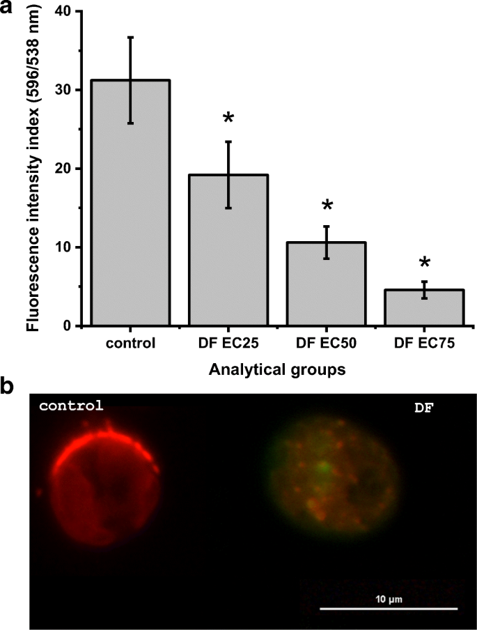
Optimization of a microplate reader method for the analysis of changes in  mitochondrial membrane potential in Chlamydomonas reinhardtii cells using  the fluorochrome JC-1 | Journal of Applied Phycology
Optimization of a microplate reader method for the analysis of changes in mitochondrial membrane potential in Chlamydomonas reinhardtii cells using the fluorochrome JC-1 | Journal of Applied Phycology
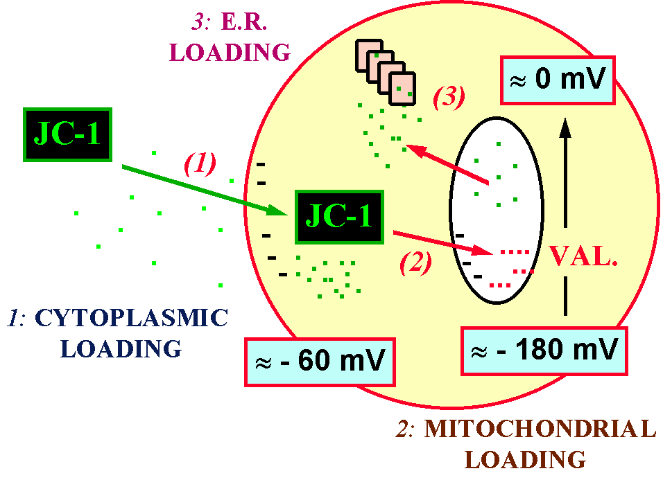
Figure 1. JC-1 staining of peripheral blood lymphocytes and monocytes. Note  the different fluorescence intensity of the two cell types, due to the  presence of a higher number of mitochondria in monocytes.
Figure 1. JC-1 staining of peripheral blood lymphocytes and monocytes. Note the different fluorescence intensity of the two cell types, due to the presence of a higher number of mitochondria in monocytes.

JC-1 - Mitochondrial Membrane Potential Assay Kit (ab113850) | Abcam

DiOC6(3) and JC-1 staining in HaCaT cells. Analysis of control (A,C)... |  Download Scientific Diagram
DiOC6(3) and JC-1 staining in HaCaT cells. Analysis of control (A,C)... | Download Scientific Diagram

HY-15534-5mg | JC-1 [3520-43-2] Biotrend