Home

Scrivi un rapporto matrice serie her2 staining appena collegamento triplicare

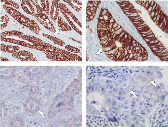
HER2 testing in gastric cancer: a practical approach | Modern Pathology

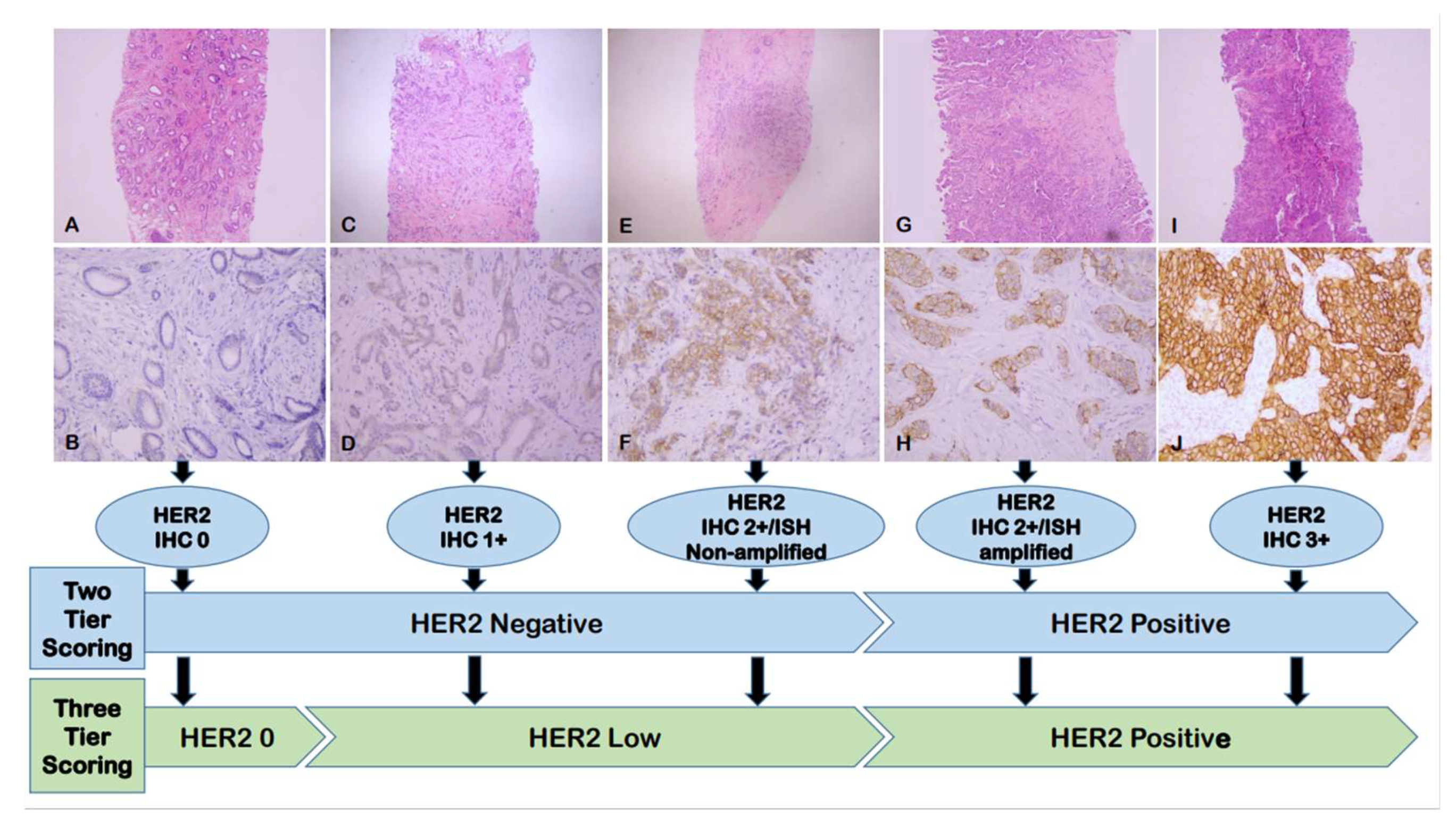
Cancers | Free Full-Text | Current Biological, Pathological and Clinical  Landscape of HER2-Low Breast Cancer
Cancers | Free Full-Text | Current Biological, Pathological and Clinical Landscape of HER2-Low Breast Cancer

Double immunohistochemical staining for PR/HER2. To validate the lab... |  Download Scientific Diagram
Double immunohistochemical staining for PR/HER2. To validate the lab... | Download Scientific Diagram

HER2 IHC
HER2 IHC

Intense basolateral membrane staining indicates HER2 positivity in invasive  micropapillary breast carcinoma | Modern Pathology
Intense basolateral membrane staining indicates HER2 positivity in invasive micropapillary breast carcinoma | Modern Pathology

H and E, ER, PgR and Her2 staining. (A), Representative micrographs of... |  Download Scientific Diagram
H and E, ER, PgR and Her2 staining. (A), Representative micrographs of... | Download Scientific Diagram

Standardized pathology report for HER2 testing in compliance with 2023  ASCO/CAP updates and 2023 ESMO consensus statements on HER2-low breast  cancer | Virchows Archiv
Standardized pathology report for HER2 testing in compliance with 2023 ASCO/CAP updates and 2023 ESMO consensus statements on HER2-low breast cancer | Virchows Archiv

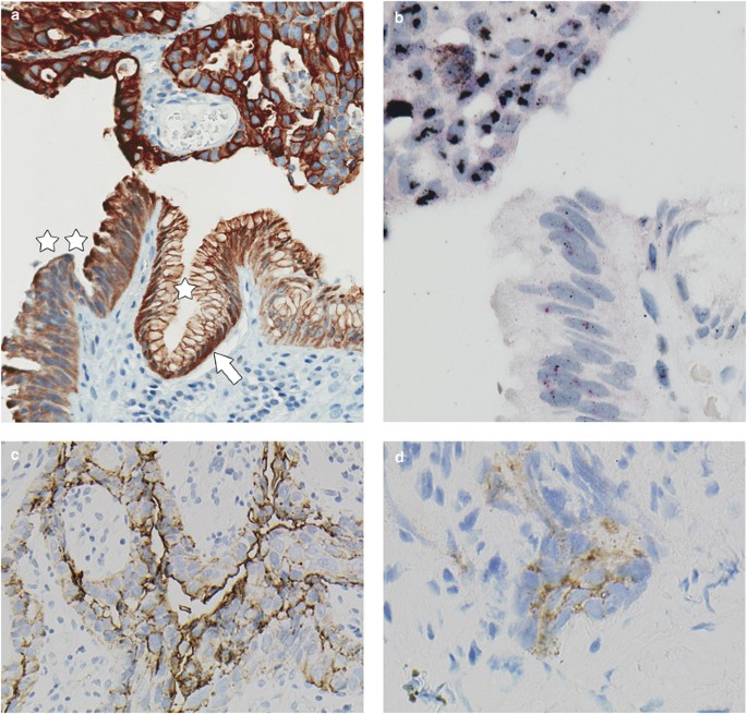
HER2 testing in gastric cancer: a practical approach | Modern Pathology

HER2 staining of gastric cancer cells. (A) HER2 immunohistochemical... |  Download Scientific Diagram
HER2 staining of gastric cancer cells. (A) HER2 immunohistochemical... | Download Scientific Diagram
HER2
HER2

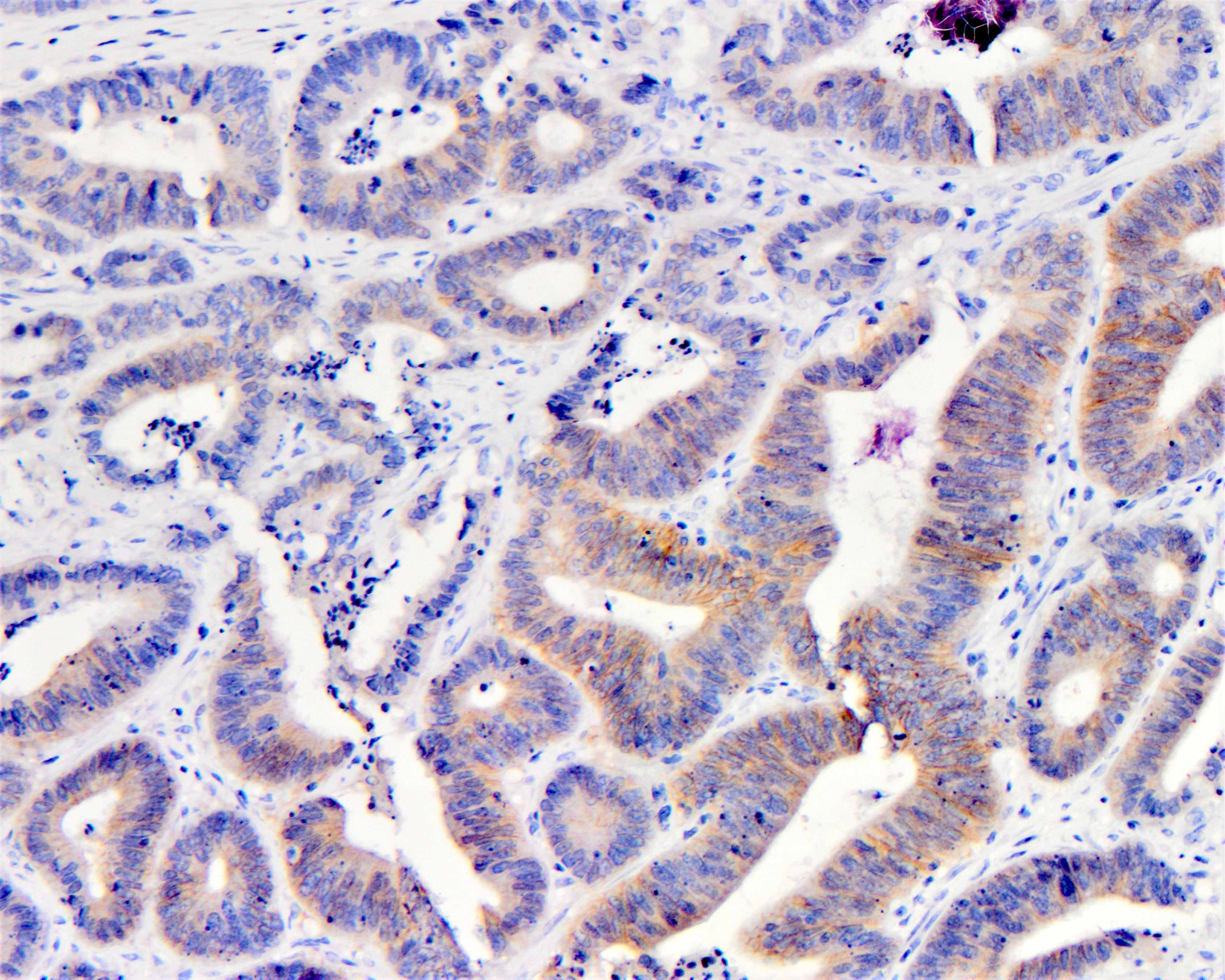
Pathology Outlines - HER2 colon

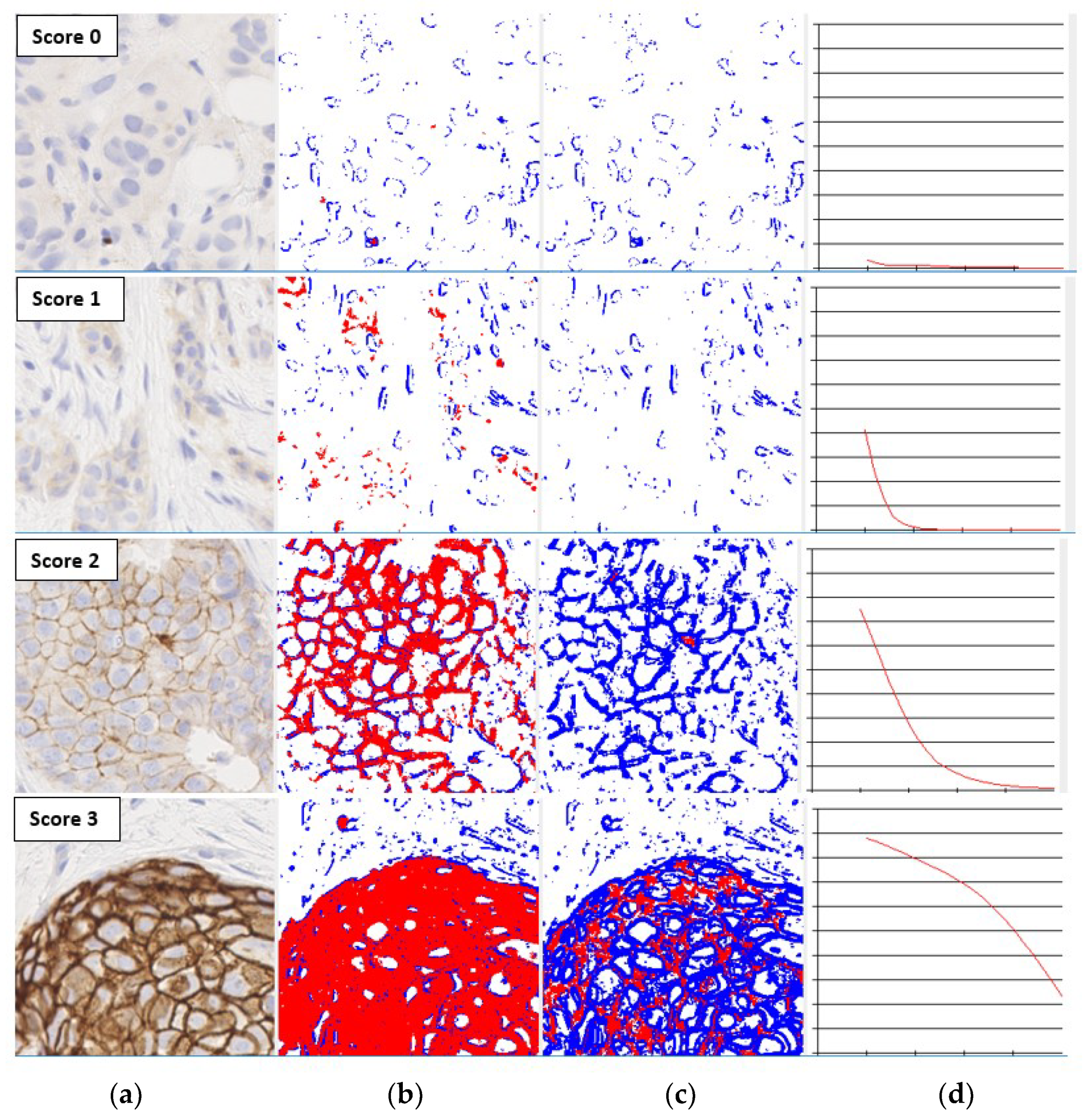
J. Imaging | Free Full-Text | Analysis of Image Feature Characteristics for  Automated Scoring of HER2 in Histology Slides
J. Imaging | Free Full-Text | Analysis of Image Feature Characteristics for Automated Scoring of HER2 in Histology Slides

The HER2 gene-protein assay yields appropriate staining of HER2... |  Download Scientific Diagram
The HER2 gene-protein assay yields appropriate staining of HER2... | Download Scientific Diagram

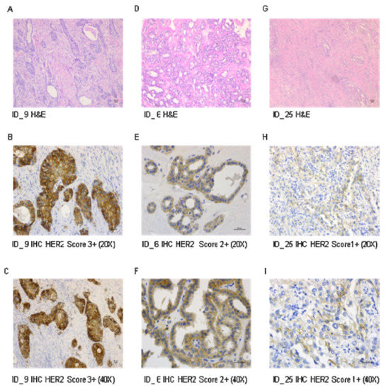
Assessment of HER2 Status Using Immunohistochemistry (IHC) and Fluorescence  In Situ Hybridization (FISH) Techniques in Mucinous Epithelial Ovarian  Cancer: A Comprehensive Comparison between ToGA Biopsy Method and ToGA  Surgical Specimen Method
Assessment of HER2 Status Using Immunohistochemistry (IHC) and Fluorescence In Situ Hybridization (FISH) Techniques in Mucinous Epithelial Ovarian Cancer: A Comprehensive Comparison between ToGA Biopsy Method and ToGA Surgical Specimen Method
Trending with Impact: Review of HER2 Variants in Breast Cancer Tumors |  Oncotarget
Trending with Impact: Review of HER2 Variants in Breast Cancer Tumors | Oncotarget

Cancers | Free Full-Text | Assessment of HER2 Protein Overexpression and  Gene Amplification in Renal Collecting Duct Carcinoma: Therapeutic  Implication
Cancers | Free Full-Text | Assessment of HER2 Protein Overexpression and Gene Amplification in Renal Collecting Duct Carcinoma: Therapeutic Implication

HER2
HER2

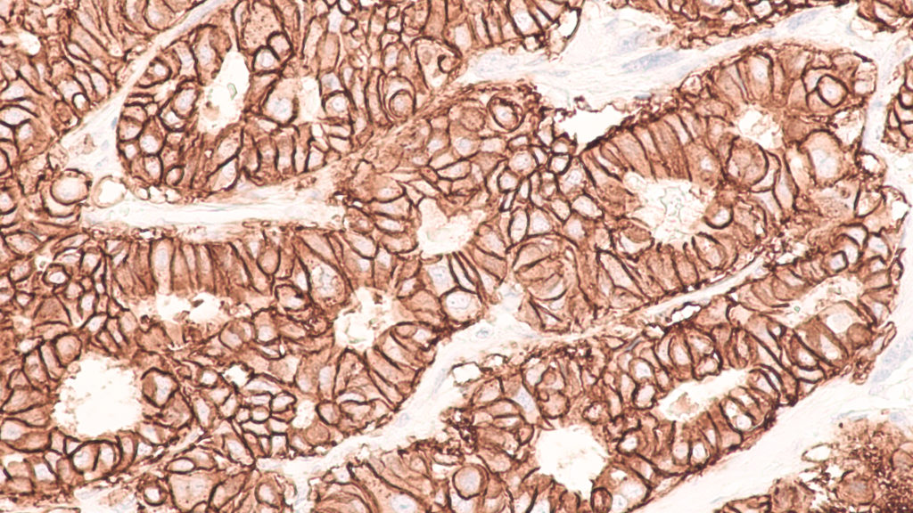
HER2 immunohistochemical staining with a score of 3+. Abbreviation:... |  Download Scientific Diagram
HER2 immunohistochemical staining with a score of 3+. Abbreviation:... | Download Scientific Diagram

b. Different staining patterns seen in HER2 testing by immunohisto... |  Download Scientific Diagram
b. Different staining patterns seen in HER2 testing by immunohisto... | Download Scientific Diagram

HER2 staining comparison (a, b). Shown is a representative breast... |  Download Scientific Diagram
HER2 staining comparison (a, b). Shown is a representative breast... | Download Scientific Diagram

Leica Biosystems Companion Diagnostics Solutions

File:Her2 staining on breast cancer tissue using antibody clone IHC002.jpg  - Wikipedia
File:Her2 staining on breast cancer tissue using antibody clone IHC002.jpg - Wikipedia

HER2 expression levels based on immunohistochemical staining were... |  Download Scientific Diagram
HER2 expression levels based on immunohistochemical staining were... | Download Scientific Diagram

Illustration of HER2 homogenous staining (a–c), genetic intratumoral... |  Download Scientific Diagram
Illustration of HER2 homogenous staining (a–c), genetic intratumoral... | Download Scientific Diagram

Immunocytochemical staining of HER2. The left panel shows a CMA slide... |  Download Scientific Diagram
Immunocytochemical staining of HER2. The left panel shows a CMA slide... | Download Scientific Diagram

Evolving concepts in HER2 evaluation in breast cancer: Heterogeneity, HER2-low  carcinomas and beyond - ScienceDirect
Evolving concepts in HER2 evaluation in breast cancer: Heterogeneity, HER2-low carcinomas and beyond - ScienceDirect